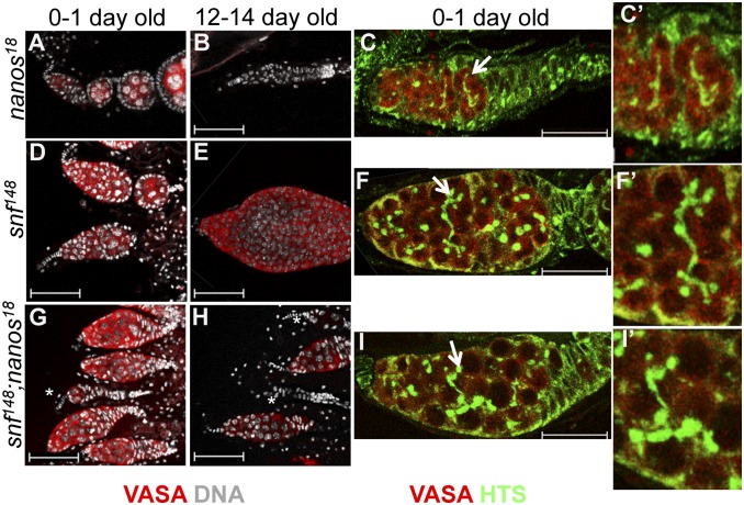
Fig. 5.
snf148, nanos18 Double mutant analysis. Comparison of germaria from (A–C) nanos18/Df(3R) Exel6183, (D–F) snf148/snf148, and (G–I) double-mutant nanos18/Df(3R)Exel6183; snf148/snf148 females stained 0–1 d or 12–14 d after eclosion for Vasa, Hu Li Tai Shao (Hts) (C, F, and I), and DNA (A, B, D, E, G, H). Arrow in C, F, and I shows the region enlarged in C', F', and I'), illustrating the difference between the thin branching fusome structure seen in nanos18/Df(3R)Exel6183 and the abnormal long branching fusome structures visible in snf148/snf148 and double-mutant nanos18/Df(3R)Exel6183; snf148/snf148 females. Scale bars: 50 μm (A, B, D, E, G, and H); 25 μm (C, F, and I). Asterisks indicate germaria with few or no germ cells.

