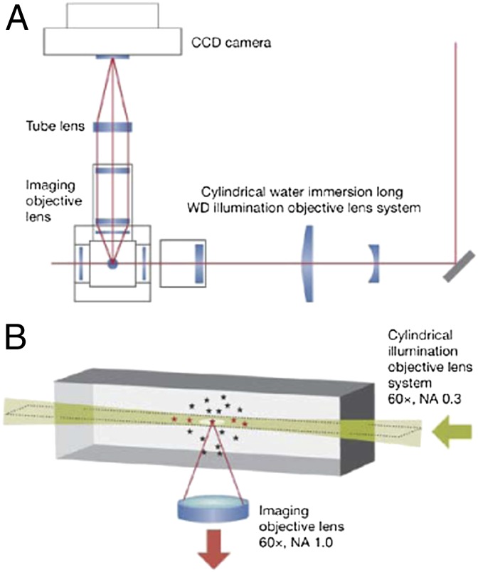
Fig. 1.
In LSFM, an excitation beam, or “sheet” (height × width × thickness, 20 μm × 20 μm × 2 μm), is delivered within the focal plane by using a cylindrical lens (magnification of 60×; NA, 0.3), and the emitted fluorescence is detected by using an objective oriented perpendicular to the illumination path (magnification of 60×; NA, 1.0). (A) Diagram of the overall microscopy system. (B) Schematic of the illumination and detection paths. CCD, cooled CCD; WD, working distance. Reproduced from ref. 16.

