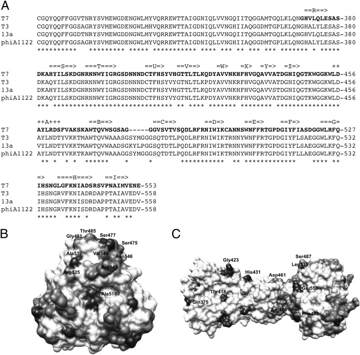
Fig. 4.
Sequence conservation of gp17. (A) Alignment of the sequence of bacteriophage T7 gp17 with its homologs from E. coli phage T3, S. enteridis phage 13a, and Y. pestis phage PhiA1122. Amino acids present in our structures are shown in bold. Residues that are identical in all four proteins are marked with asterisks. Secondary structure elements identified in our structure are also indicated, beta-strands with arrows and α-helices with plus-signs. (B and C) Sequence conservation mapped on the structure. The color scale is from white (absolutely conserved) to black (no conservation). A top view (B) and a side view (C) are shown. Residues that are not conserved, and thus may be important for host range discrimination, are indicated.

