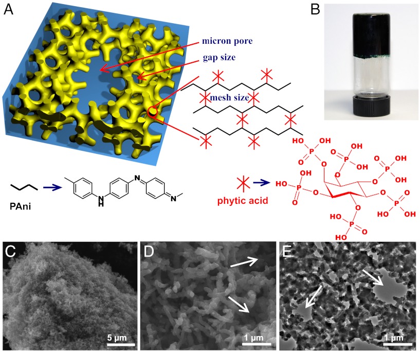
Fig. 1.
Chemical structure and morphological characterization of phytic acid gelated and doped polyaniline hydrogel. (A) Schematic illustrations of the 3D hierarchical microstructure of the gelated PAni hydrogel where phytic acid plays the role as a dopant and a crosslinker. Three levels of hierarchical porosity from angstrom, nanometer to micron size pores have been highlighted by red arrows. (B) A photograph of the PAni hydrogel inside a glass vial. (C) SEM image of a piece of dehydrated hydrogel. (D) A higher magnification SEM image showing the interconnected network of dendritic PAni nanofibers. (E) A TEM image showing the continuous nanostructured network of the dehydrated PAni hydrogel. The white arrows in D and E denote the micron size pores in PAni hydrogel.

