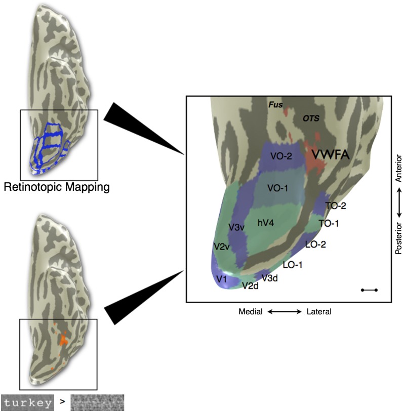
Fig. 1.
Location of VWFA in relation to the visual field maps in a typical subject. (Upper Left) fMRI retinotopic mapping was performed to define visual field map boundaries (blue) for V1, V2, V3, hV4, VO-1, VO-2, LO-1, LO-2, TO-1, and TO-2. (Lower Left) Separate runs of the VWFA localizer were performed, and voxels were chosen on the basis of the contrast words > phase-scrambled words at a threshold of P < 0.001 (uncorrected, orange overlay). VWFA voxels were restricted to be outside of known retinotopic areas and anterior to hV4 in ventral occipito-temporal cortex. (Right) The cortical surface of the posterior left hemisphere is rendered as a mesh and computationally inflated to visualize the sulci (dark gray). The majority of VWFA voxels (red) are located lateral or anterior to VO-1/VO-2 in all subjects. The TO-1/TO-2 maps overlap with the human motion complex (hMT+). Fus, fusiform gyrus; OTS, lateral occipitotemporal sulcus. (Scale bar: 10 mm.) Subject is S1.

