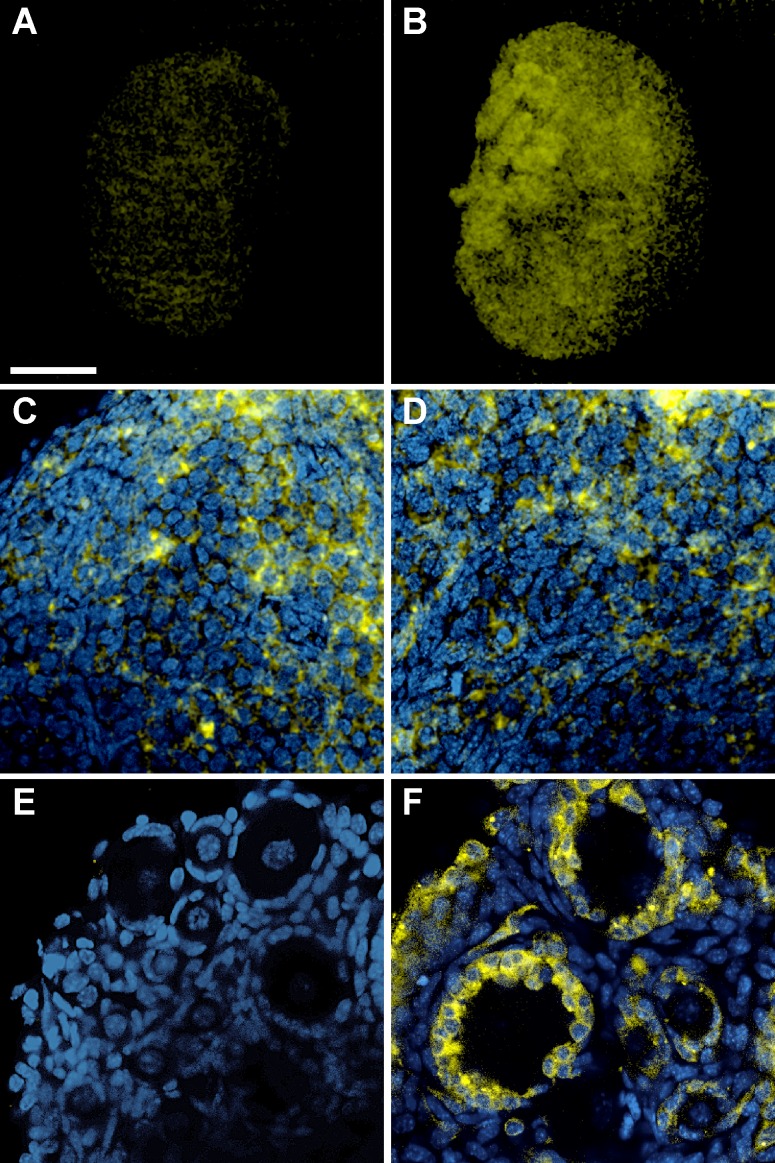
FIG. 1. .
Expression of the SMOM2/YFP fusion protein examined by confocal microscopy of whole-mount ovaries from Amhr2+/+SmoM2 control and Amhr2cre/+SmoM2 mutant mice on the day of birth (A–D) and at Day 10 of age (E, F). Images of control ovaries are shown in A and E and all other panels show images of mutant ovaries. C and D show higher-power images of cortex and medulla, respectively, of the ovary shown in B. Bar in A = 200 μm for A and B and 40 μm for C–F.

