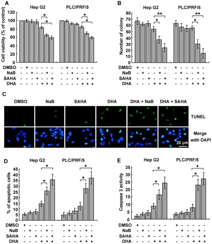
Figure 3. HDACi facilitated DHA-induced apoptosis. A.
Combination treatment with HDACi and DHA increased cell death. Cells were treated with 4 mM NaB, 1.25 μM SAHA, 10 μM DHA or combination of NaB/SAHA and DHA for 24 h. Cell viabilities were measured by MTT assay. B. The inhibitory effect of HDACi and DHA in combination on liver cancer cell growth was further confirmed by colony formation assay. One hundred of cells were seeded into 6-well plates for 7 d, and then cultured with either HDACi or DHA for another 7 d. Colonies were stained with 0.05% crystal violet. The number of colony in each well was counted and statistical analysis was performed. Data are presented as mean ± SD of three independent experiments. C. The effect of HDACi on DHA-induced apoptosis was measured by TUNEL assay, using in situ cell death detection kit. Hep G2 cells treated as described in A were subjected to TUNEL assay. Apoptotic cells were observed under fluorescent microscope. D. The effect of HDACi and DHA in combination was further confirmed by TUNEL assay, using flow cytometry. Percentage of apoptotic cells was calculated. E. Caspase 3 activation was involved in HDACi-mediated apoptosis in cells treated with DHA. The activity of caspase 3 in cells treated as described in A was determined and the relevant change was shown. For A, B, D and E, *P<0.05, **P<0.01, versus the DHA group.

