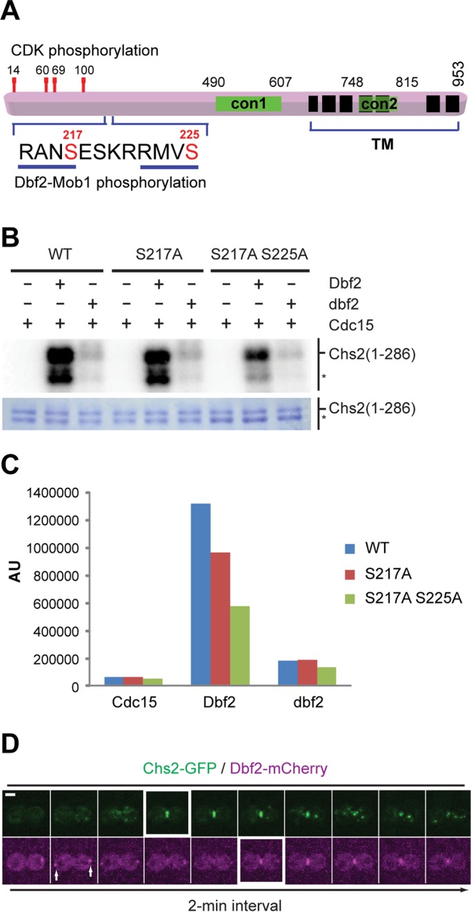
FIGURE 1:

Mitotic exit kinase Dbf2 directly phosphorylates Chs2. (A) Schematic diagram of Chs2. con1 and con2, conserved regions of typical chitin synthases; TM, predicted transmembrane domains. Demonstrated and putative phosphorylation sites for CDK1 and Dbf2 kinases in the N‑terminal region of Chs2 are indicated. (B) In vitro phosphorylation of the N‑terminal region of Chs2 by Dbf2. Asterisk, degradation product of GST-Chs2 (1–286). (C) Quantification of Dbf2-mediated phosphorylation of Chs2. (D) Time-lapse analysis of a yeast cell coexpressing Chs2-GFP and Dbf2-mCherry (YO1182). Cells were grown in SC-HIS media to exponential phase at 25°C and then processed for time‑lapse microscopy. Arrows, spindle pole bodies. Scale bar, 2 μm.
