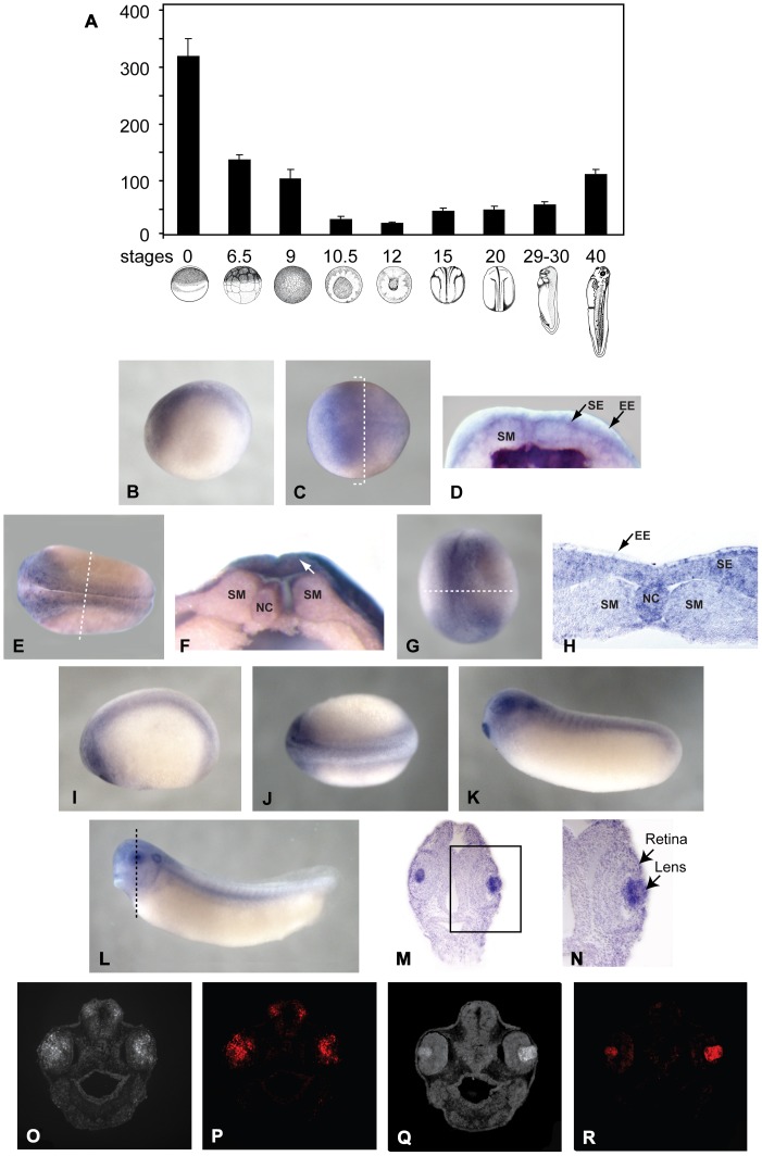
Figure 2. Expression of SPC7 mRNA in Xenopus embryos at different developmental stages.
(A) Real-time PCR. (B-N) Whole mount hybridization in situ (in panels B, C, E, and I-L, anterior is to the left. Dotted lines in C, E, G, and L indicate the approximate plane for images in D, F, H, and M, respectively). (B-C) Lateral (B) and dorsal (C) views of a stage 13 embryo showing staining in the early neural plate. (D) Hemisected stage 13 embryo displaying detectable expression of SPC7 in the sensorial layer of the neuroectoderm, but not in the underlying somitogenic mesoderm. (E) Dorsal view of stage 16 embryo showing SPC7 staining in the neural folds and presumptive eye field. (F) hemisected midneurula (stage 16) showing staining in the sensorial ectoderm (arrow). (G) Antero-dorsal view of a late neural fold (stage 18) embryo showing staining in the anterior part of neural plate and anterolateral edges. (H) Plastic section through over-stained embryo showing staining in neuroectoderm. Lateral (I) and dorsal (J) views of a stage 21–22 embryo showing prominent staining in the eye, cement gland, brain, and neural tube. (K) Lateral view of stage 26 embryo showing SPC7 expression in the retina, lens, cement gland, otic vesicle, and throughout the head and somites. (L) At stage 32, staining in the lens became more prominent, primarily because expression in the retina decreased substantially. (M) Plastic section of over-stained stage 35 embryo showing specific SPC7 localization in the lens. (N) Enlarged view of boxed area shown in M. (O-R) Stage 30–32 (O, P) and stage 35–37 (Q, R) paraffin sections analyzed for SPC7. (O) Total fluorescence. (P) Same section as in O showing specific SPC7 staining in retina and lens (Fast Red). (Q) Total fluorescence. (R) Same section as in Q showing specific SPC7 fluorescent staining localized to the lens. (NC = Notochord; EE = Epithelial Layer of Neuroectoderm; SE = Sensorial Layer of Neuroectoderm; SM = Somitogenic Mesoderm).

