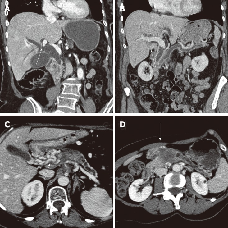
Figure 1.

Ductal dilation, computer tomography 3-phase contrast-enhanced thin-slice helical scan. A: Heterogenous tumor of the pancreatic head with consecutive extra- and intrahepatic bile duct dilatation (arrow); B: “Double duct sign” due to a tumor of the papilla of vater (arrow); C: Tumor of the pancreatic neck with an upstream dilatation of the pancreatic duct and parenchymal atrophy of the pancreatic gland. Presence of a cavernoma due to tumor thrombosis of the portal vein (arrow); D: Classic radiological presentation of a pancreatic neck tumor with a less pronounced enhancement compared to the normal pancreatic parenchyma (arrow).
