Figure 1.

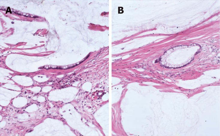
Peritoneal lesions consist of scant strips (A) and gland (B) of histologically bland mucinous epithelium associated with abundant extracellular mucin and fibrousis in disseminated peritoneal adenomucinosis (HE, × 40).

Peritoneal lesions consist of scant strips (A) and gland (B) of histologically bland mucinous epithelium associated with abundant extracellular mucin and fibrousis in disseminated peritoneal adenomucinosis (HE, × 40).