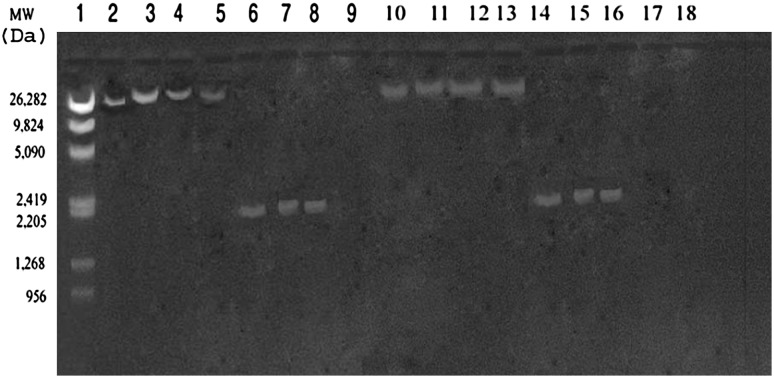
Fig. 1.
Agarose gel (0.8%) showing 26 and 2 Kb plasmids in donors (Lane 2–8) and their respective transconjugants (Lane 10–16). Molecular weight marker λ/Mlu Hind digest along with their size (in Daltons) is shown in lane 1. The recipient strain (E. coli K12F−SR lac−) screened for plasmids before transconjugation experiment was found devoid of the same (lane 9)

