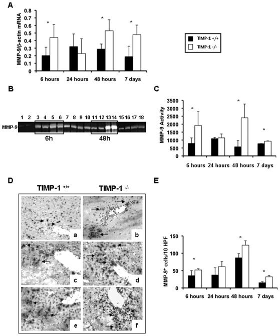
Figure 4.
MMP-9 expression and activity in TIMP-1−/− and TIMP-1+/+ mice. MMP-9 mRNA expression (panel A), as detected by RT-PCR analysis, was significantly upregulated in TIMP-1−/− mice at 6h, 48h, and 7d after IR injury, as compared to the respective wild-type controls. MMP-9 activity (panels B and C), analyzed by zymography in TIMP-1+/+ (lanes 1, 3, 4, 7, 8, 11, 12, 15, and 16) and TIMP-1 −/− (lanes 2, 5, 6, 9, 10, 13, 14, 17, and 18) livers; MMP-9 activity was almost absent in naïve livers of TIMP-1+/+ (lane 1) and TIMP-1−/− (lane 2) mice and highly detectable in TIMP-1+/+ and TIMP-1−/− livers at 6h (lanes 3-6), 24h (lanes 7-10), 48h (lanes 11-14), and 7d (lanes 15-18) post-IRI; however, compared to controls, MMP-9 activity was markedly upregulated in TIMP-1−/− livers at 6h, 48h, and 7d after IRI. MMP-9+ cells (panel D and E) in wild-type controls (a, c, e) and TIMP-1−/− livers (b, d and f) at 6h (a, and b), 24h (c, and d), and 48h (e, and f) post-IRI; MMP-9+ cells were detected in significantly higher numbers in TIMP-1−/− livers, particularly at 6h, 48h, and 7d post-reperfusion, (n=4-5/group; *p<0.05).

