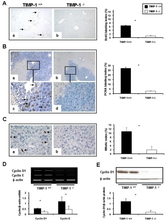
Figure 6.
Expression of hepatic regenerative markers in TIMP-1−/− and TIMP-1+/+ mice. Hepatocyte BrdU incorporation (panel A), PCNA labeling (panel B), and phosphorylated histone H3-positive cells (panel C) in TIMP-1+/+ (a, and c) and TIMP-1−/− (b, and d) livers at 48h post-IRI; PCNA staining (c, and d) is shown in higher magnification to better illustrate positive (c) and virtually negative (d) PCNA hepatocyte-labeling in the surviving parenchyma of TIMP-1+/+ and TIMP-1−/− livers, respectively. TIMP-1−/− livers showed markedly diminished BrdU, PCNA, and mitotic labeling indexes, as compared to controls. The densitometric ratios of cyclin D1/β-actin and cyclin E/β-actin mRNA (panel D) were significantly depressed in TIMP-1−/− livers at 48h post-IRI. Cyclin D1 at protein level (panel E) was also profoundly depressed in TIMP-1−/− livers at 48h post-IRI, (n=4-5/group; *p<0.05).

