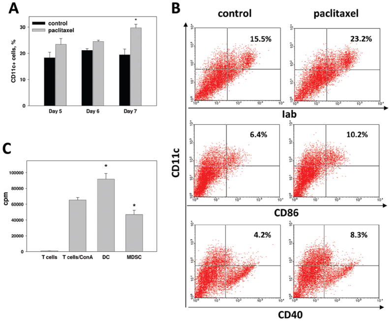
Figure 4.
Paclitaxel stimulates differentiation of MDSC into DC. (a) MDSC cultures were treated with 1 nM paclitaxel and the amount of CD11c+ DC was assessed after 24–72 h by FACScan, as described in Materials and methods. Results from four independent experiments are shown as the mean (± SEM). (b) Additional phenotyping of DC revealed that they express MHC Class II molecules and low levels of CD86 and CD40. (c) CD11c+ DC were isolated from paclitaxel-treated MDSC cultures using magnetic beads (DC) and co-cultured with ConA-activated syngeneic splenocytes (T-cells/ConA). DC-depleted MDSC served as a control (MDSC). T-cell proliferation was assessed by [3H]-thymidine incorporation and express as count per minute (cpm). * p < 0.05 vs corresponding control (a) or ConA-stimulated T-cell proliferation (c). (See colour version of this figure online at www.informahealthcare.com/imt)

