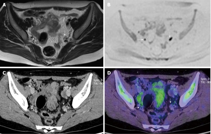
Figure 2.

A 51-year-old woman with cervical cancer with lymph node metastases in left internal iliac area. A: T2-weighted magnetic resonance imaging shows two small lymph nodes (LNs) in right and left internal iliac areas (arrows); B: These two LNs seen in (A) show moderately abnormal signal intensity on diffusion-weighted imaging (DWI) (arrows), suggesting the presence of nodal cancer spread; C: Enhanced computed tomography (CT) component of positron emission tomography and CT (PET/CT) shows two small LNs in right and left internal iliac areas (arrows); D: PET/CT shows no 18F-fluorodeoxyglucose uptake corresponding to the two LNs seen in (C) (arrows) suggesting the absence of nodal cancer spread. Histopathological specimen findings confirmed extensive LN involvement by cancer in left internal iliac LN and no involvement in right internal iliac LN. DWI was false-positive for the right and true-positive for the left node. PET/CT was true-negative for the right and false-negative for the left node.
