Fig. 2.
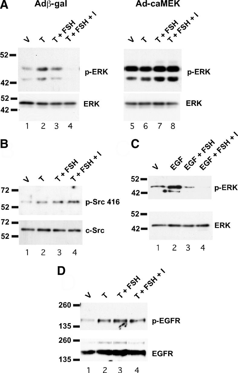
FSH acts upstream of MEK and downstream of the EGF receptor to inhibit ERK activity. A, Wild-type Sertoli cells were transfected with adenoviral constructs expressing either β-galactosidase (Adβ-gal) or constitutively active MEK (Ad-caMEK) for 2 d and then were stimulated with EtOH-vehicle (V), 100 nm testosterone (T), T + FSH (100 ng/ml), or T + FSH + IBMX (I, 0.5 mm) for 10 min. Whole-cell extracts were assayed for p-ERK and total ERK levels by Western blot. B, Wild-type Sertoli cells were treated with V, T, or T + various combinations of FSH or IBMX for 10 min. Whole-cell extracts were assayed by Western blot using antisera against Src phosphorylated at position 416 or total Src. C, Wild-type Sertoli cells starved of EGF for 16 h were stimulated with V, EGF (10 nm), or EGF + FSH, or EGF + FSH + IBMX (B). Whole-cell extracts were assayed for p-ERK, and total ERK levels were assayed by Western blot. D, Wild-type Sertoli cells were treated as in A and whole-cell extracts were immunoprecipitated with antiserum against total EGFR and then assayed for EGFR phosphorylated at position 1173 and total EGFR by Western blot. The blots shown are representative of three independent experiments.

