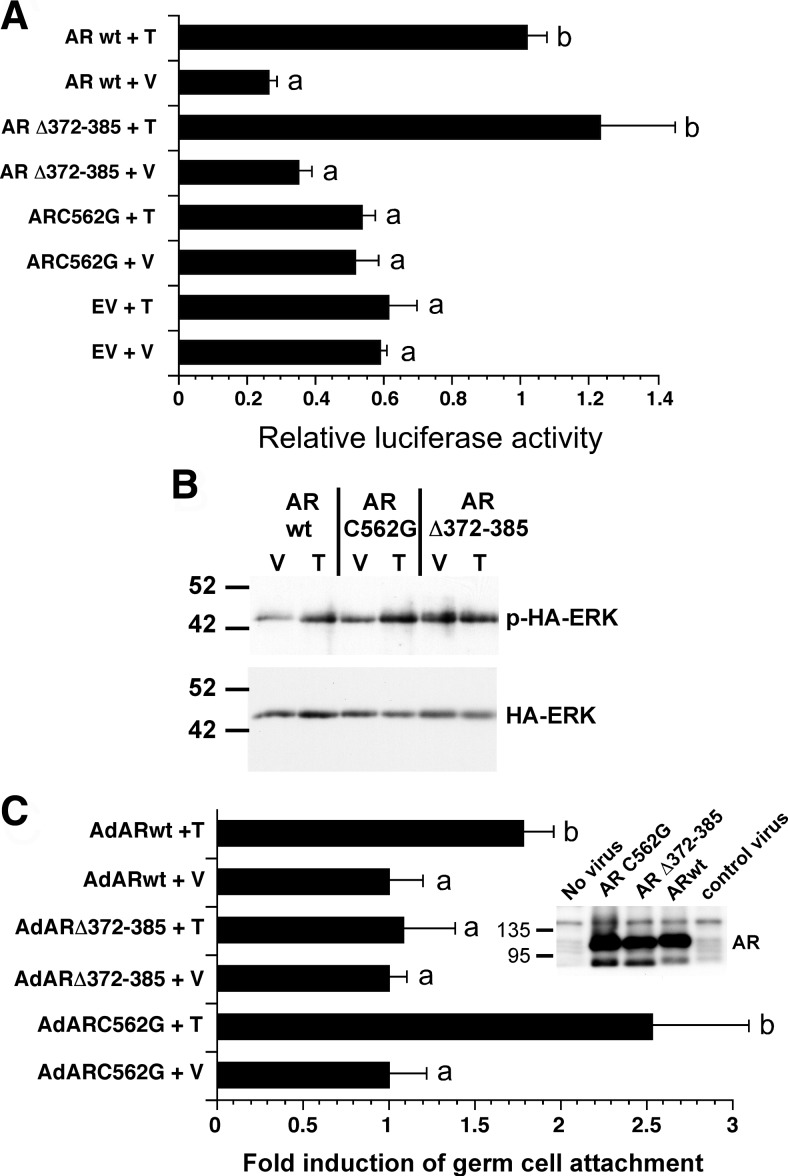
Fig. 5.
The nonclassical pathway contributes to testosterone-mediated Sertoli-germ cell attachment. A, Sertoli cells isolated from 15-d-old tfm rats were transfected with empty vector (EV) or vectors expressing the ARC562G or ARΔ372–385 mutant forms of AR or the wild-type AR (AR wt) as well as a luciferase reporter plasmid driven by the AR-inducible PSA promoter. The cells were stimulated with EtOH vehicle (V) or 100 nm testosterone (T) for 24 h, and luciferase activity was determined after normalization for protein content. For each AR construct, the mean (±se) relative luciferase activity is normalized to that of ARΔ wt + T (=1). Values with different lowercase letters differ significantly (P < 0.05) (n = 3). B, Sertoli cells isolated from tfm rats were cotransfected with plasmids encoding wild-type AR, ARC562G, or ARΔ372–385 and a plasmid encoding HA epitope-tagged ERK. After stimulation with V or T for 10 min, whole-cell extracts were immunoprecipitated with antiserum against the HA epitope followed by Western blot analysis using an antiserum against phosphorylated ERK or total ERK. The image shown is representative of three experiments. C, Sertoli cells isolated from 15-d-old tfm rats were infected with adenovirus constructs expressing wild-type AR, ARΔ372–385, or ARC562G. germ cells were added 2 d later in the presence of vehicle (V) or 100 mm testosterone (T), and the number of germ cells attached per Sertoli cell was determined 48 h later. For each adenovirus construct, the mean (±se) percentage of germ cells bound per Sertoli cell was normalized to that of vehicle treatment (=1), and the results of testosterone treatment were expressed as fold induction over the paired vehicle levels. For comparisons of treatments for individual adenovirus constructs, values with different lowercase letters differ significantly (P < 0.05) (n = 4). Cells were counted from at least five random fields for each of three studies. The inset shows the results of a Western blot using extracts from COS-7 cells infected with no virus, AdARC562G, AdARΔ372–385, AdARwt, and AdRAP1DN (control virus).

