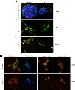
Fig. 4.
MafA is selectively reduced in the β-cells of Hnf1α−/− mice. A, MafA, insulin, and CK19 expression were examined by immunofluorescence in pancreatic sections from 3-month-old adult Hnf1α−/− and wild-type control mice. B and C, MafA expression was also compromised in E17.5 and E18.5 Hnf1α−/− insulin+ cells, whereas little-to-no apparent change was observed in insulin (B and C), Nkx2.2, Pdx1, Nkx6.1, and Pax6 (D) levels compared with control. White arrowheads point to MafA+ nuclei in the WT sections, whereas the yellow arrowheads illustrate the MafA− nuclei within Hnf1α−/− insulin+ cells. Representative images are shown for each staining condition. Notably, the reduction in MafA levels was observed in embryonic as well as adult Hnf1α−/− samples.

