Fig. 1.
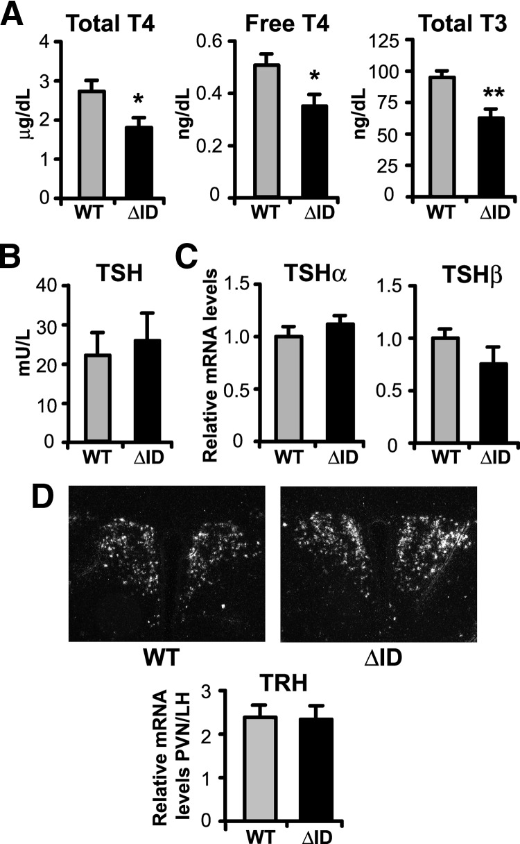
NCoRΔID mice have reduced circulating TH levels accompanied by normal serum TSH and normal TSH and TRH mRNA expression. A, Serum total T4, free T4, and total T3 were measured by RIA in male adult WT and NCoRΔID mice (n = 6–10 per group). B, Serum TSH was measured in the same groups of animals by high-affinity RIA (n = 6–7 per group). C, TSH subunit mRNA levels in pituitary of male adult mice were quantified by quantitative PCR. mRNA levels are expressed relative to WT group (n = 7 animals per group). D, ISH was performed on formalin-fixed brain sections from male mice with indicated genotypes using a 35S-labeled riboprobe against mouse TRH mRNA. Shown are representative images of the PVH at original magnification, ×10. The TRH mRNA expression was quantified using Image J software. Data are presented as relative pixel densities in PVH normalized to lateral hypothalamus (LH) (n = 5 animals per group). Data are presented as mean ± sem. *, P < 0.05; **, P < 0.01.

