Fig. 6.
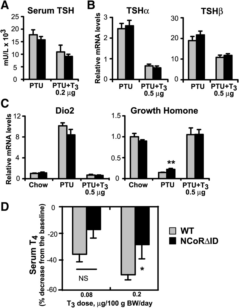
The HPT axis of NCoRΔID mice responds normally to changes in serum TH level. A, Serum TSH was measured by high-affinity RIA in hypothyroid (PTU) and low T3 replacement dose groups (n = 3–7 animals per group). B, TSH subunit mRNA levels were quantified by quantitative PCR in hypothyroid and physiological T3 replacement dose groups. mRNA levels are expressed relative to WT euthyroid animals (set to 1) (n = 5–7 animals per group). C, Expression of known TH targets in the pituitary of NCoRΔID mice at different circulating T3 levels. mRNA expression levels were quantified by quantitative PCR and presented relative to euthyroid WT group (set to 1) (n = 5–7 animals per group). D, Feedback suppression of thyroid function by exogenous T3. WT and NCoRΔID mice were given injections of two indicated incremental doses of T3 for 4 d each. Total serum T4 levels were measured at the end of each treatment period, and the decrease was expressed as a percentage of the baseline obtained in the same animals before the treatment (n = 7–9 animals per group). Data are presented as mean ± sem; * P < 0.05; **P < 0.01.

