Fig. 2.
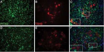
Efficient deletion of Pitx2 in Pitx2flox/−;Tg(Tshb-cre) mice. Paraformaldehyde-fixed, paraffin-embedded pituitary glands from 8-wk-old control (A–C) and Pitx2flox/−;Tg(Tshb-cre) mice (D–F) were sectioned and coimmunostained with PITX2 (green, panels A and D) and TSH (red, panels B and C), counterstained with DAPI (blue) and photographed, and images were merged (C and F) (×630). The majority of thyrotroph cells express PITX2 in controls (panel C) but not in Pitx2flox/−;Tg(Tshb-cre) mice (panel F). Boxed areas are magnified in the inset. The magnification bar in the TSHB and PITX2 immunostaining is equivalent to 10 μm and applies to all the pituitary hormone antibody staining.

