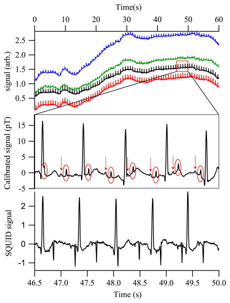
Fig. 2.
(Color online) (Top) Real-time MCG from a 31 week fetus, showing all magnetometer channels. (Middle) Portion of channel 2, with an 80 Hz low-pass filter and a 60 Hz comb filter applied. The fetal QRS complexes are circled; arrows identify the fetal P-wave components. (Bottom) SQUID gradiometer signal with the same filters applied. The gradiometry suppresses the maternal MCG as compared to the fMCG.

