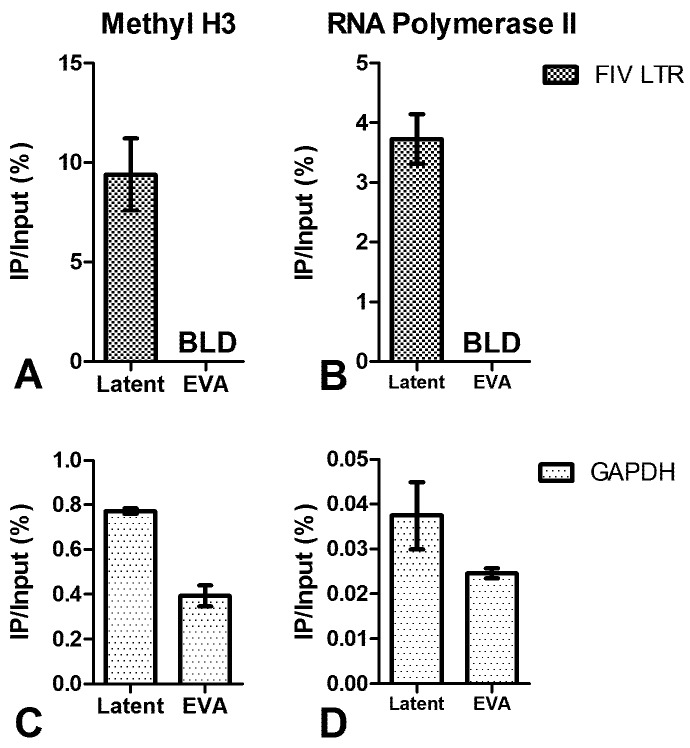
Figure 5.
ChIP assay of FIV-infected CD4+ T-cells using anti-methylated histone H3 and anti-RNA Polymerase II antibodies. (A) The FIV promoter of latently-infected, but not ex vivo activated (EVA) [5], CD4+ T-cells is associated with methylated H3; (B) The FIV promoter of latently-infected, but not ex vivo activated (EVA), CD4+ T-cells is associated with RNA Polymerase II; (C,D) GAPDH positive controls are shown for A and B respectively. Data are presented as the percentage of immunoprecipitated (IP) DNA out of the total input DNA with background subtraction using a normal IgG control. Error bars represent the standard deviation of triplicate qPCR measurements, and data is representative of ChIP experiments repeated in two different cats (A/C: cat #184 and #187, B/D: cat #165 and #187) between 29 and 32 months post FIV infection.

