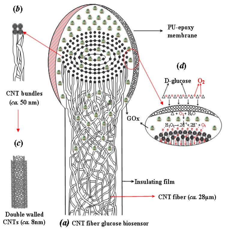
Figure 5.
Schematic diagram showing (a) CNT fiber based glucose biosensor, (b) CNT bundles, (c) DWNT and (d) working principle of biosensor. The CNT fiber (ca. 28 μm) is made of bundles (ca. ∼50 nm) of DWNTs (ca. 8–10 nm) entangled to form concentrically compacted multiple layers of nano-yarns along the CNT fiber axis, as illustrated in (a). GOx (GOD) enzyme is immobilized at the brush-like end of the CNT fiber and the enzyme layer is encapsulated by the epoxy-polyurethane (EPU) semi-permeable membrane [68].

