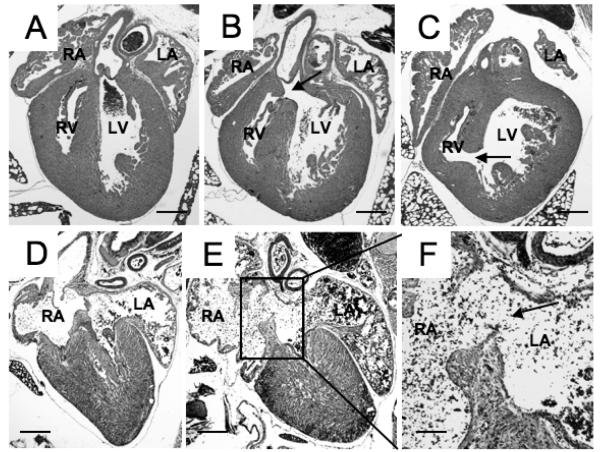
Figure. 3. A variety of septal defects were observed in mutant and trisomic mice at P0.
A) Normal heart showing intact ventricular septum at P0; B) membranous VSD; C) muscular VSD ; D) normal heart showing atrial septum; E) ostium secundum ASD; F) ASD at higher magnification. For the incidence of defects in various models, see Table 2. Arrows indicate communication between the chambers. RA: right ventricle; LV: left ventricle; RA: right atrium; LA: left atrium; Scale bars: A-E, 400 μm, f, 150 μm.

