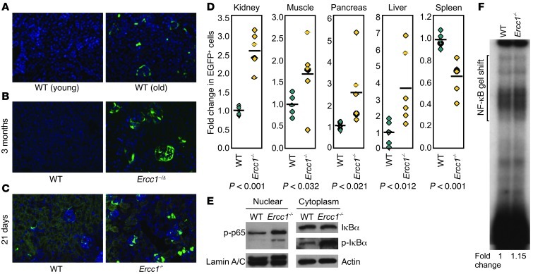
Figure 1. NF-κB activation is increased in tissues of old WT and progeroid, DNA repair–deficient mice.
Kidney sections from NF-κBEGFP mice were imaged using fluorescent microscopy to detect EGFP expression (green). Nuclei were counterstained with Hoechst dye (blue; original magnification, ×20). (A) Young adult (3-month-old) and old WT NF-κBEGFP (2-year-old) mice. (B) Ercc1–/ΔNF-κBEGFP and WT NF-κBEGFP mice at 3 months of age. (C) Ercc1–/–NF-κBEGFP and WT NF-κBEGFP mice at 21 days of age. (D) Quantification of EGFP expression. The number of EGFP+ cells was counted in 5 random fields of tissue per mouse (n = 6 mice per group). The fold difference in the number of EGFP+ cells relative to the mean value (black bar) of the group is reported. Diamond symbols represent individual mice (controls in green and Ercc1–/–NF-κBEGFP mice in yellow). P values were calculated using a Student’s t test. (E) Ercc1–/– and WT primary MEFs were passaged 5 times at 20% O2 to promote the onset of senescence (58). The levels of p-p65, IκBα, and p-IκBα in nuclear and cytoplasmic extracts were measured by immunoblot. (F) NF-κB EMSA was performed with a radiolabeled oligonucleotide containing an NF-κB binding site using nuclear extracts from Ercc1–/– and WT primary MEFs.

