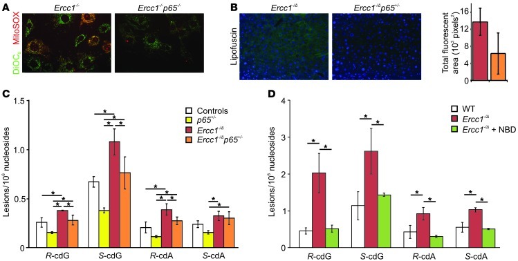
Figure 6. Inhibition of NF-κB reduces oxidative stress and damage in vitro and in vivo.
(A) Ercc1–/– and Ercc1–/–p65–/– passage 6 primary MEFs grown at 20% O2 were stained with DiOC6 (green) to mark mitochondria and MitoSOX (red) to detect mitochondrial superoxide anion (original magnification, ×40). (B) Liver sections from 10-week-old Ercc1–/Δ and Ercc1–/Δp65+/– mice imaged for lipofuscin fluorescence (original magnification, ×20). The histogram indicates the total fluorescent area for 5 images from 3 different mice per genotype calculated using MetaMorph software. (C) The levels of the (5′R) and (5′S) diastereomers of cdG and cdA in nuclear DNA isolated from the livers of 10-week-old control, p65+/–, Ercc1–/Δ, and Ercc1–/Δp65+/– mice. (D) The levels of cdG and cdA in nuclear DNA isolated from the livers of 19-week-old control, untreated Ercc1–/Δ, and 8K-NBD–treated Ercc1–/Δ mice. *P < 0.05, Tukey-Kramer test. Values denote the mean ± SEM (n = 3 per group).

