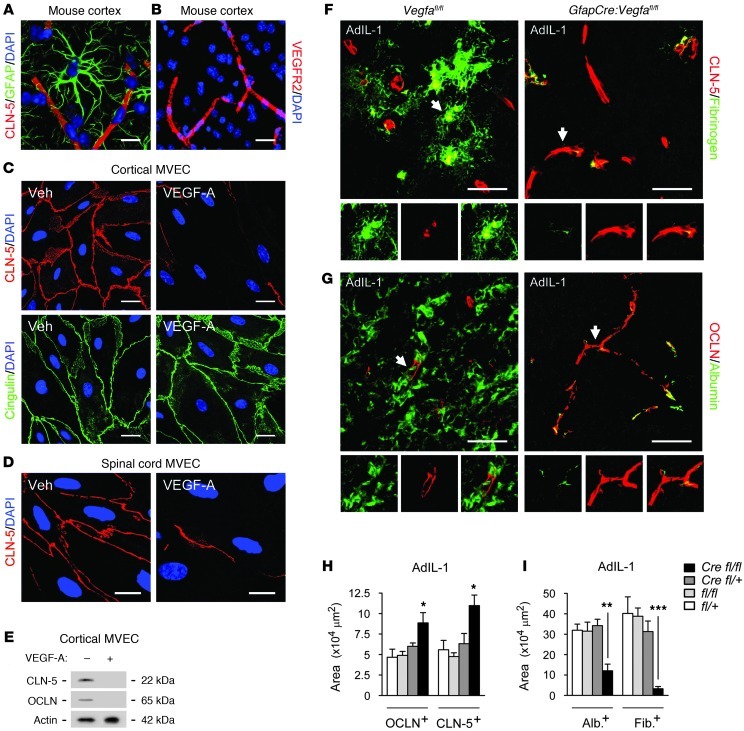
Figure 2. GfapCre:Vegfafl/fl animals display reduced BBB breakdown.
(A) 3-dimensionally rendered projection from cortex of a 12-week-old C57BL/6 mouse, illustrating intimate association of GFAP+ astrocytic endfeet with CLN-5+ endothelium. (B) Cortical section from the same mouse immunostained for VEGFR2, demonstrating localization to endothelium. (C–E) MVEC cultures from mouse cortex (C and E) and spinal cord (D) were treated with vehicle or 10 ng/ml VEGF-A and harvested at 24 hours, followed by immunostaining (C and D) and immunoblotting (E). Results are typical of data from 3 separate cultures. (F–I) AdIL-1–injected cerebral cortices from 12-week-old GfapCre:Vegfafl/fl mice and littermates sacrificed at 7 dpi (n = 21, at least 4 per genotype, as in Figure 1). (F and G) Immunostaining for CLN-5 (F) and OCLN (G). Individual channels from sections of the merged images are shown below, enlarged 1.5-fold. (H) Morphometry of OCLN and CLN-5. (I) Morphometry of albumin and fibrinogen extravasation (measures of BBB breakdown). Data are representative of 3 independent experiments. Scale bars: 20 μm (A and C); 40 μm (B); 10 μm (D); 50 μm (F and G). *P < 0.05, **P < 0.01, ***P < 0.001, ANOVA plus Bonferroni test.

