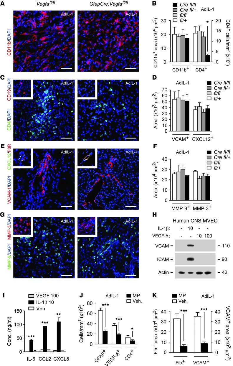
Figure 3. Reduced lymphocyte infiltration in GfapCre:Vegfafl/fl mice.
(A–G) AdIL-1–microinjected cortices were harvested at 7 dpi from 12-week-old GfapCre:Vegfafl/fl mice and littermates (at least 4 per genotype, n = 21). Shown are immunostaining and morphometry of (A and B) CD11b, (B and C) CD4, (C) CD19, (D and E) VCAM-1 and CXCL12, and (F and G) MMP-9 and MMP-3. Data are representative of findings from 3 independent experiments. (H and I) Human CNS MVECs were treated with 10 ng/ml IL-1β or 10 or 100 ng/ml VEGF-A for 24 hours, and (H) expression of VCAM-1 and ICAM-1 were determined by immunoblotting, and (I) concentrations of CC and CXC chemokines and cytokines were quantified. Data are representative of 3 experiments in separate cultures. (J and K) 12-week-old C57BL/6 mice (n = 4 per group) received 50 mg/ml MP or vehicle i.p., then intracerebral microinjection of AdIL-1 24 hours later, and were sacrificed at 7 dpi and examined to determine (J) the proportion of GFAP+, VEGF-A+, and CD4+ cells (measures of immunoreactivity and lymphocyte infiltration) and (K) the expression of fibrinogen and VCAM (measures of BBB breakdown). Data are representative of findings from 3 independent experiments. Scale bars: 50 μm (A, C, E, G, and all insets). *P < 0.05, **P < 0.01, ***P < 0.001, ANOVA plus Bonferroni test (B, D, F, and I) or Student’s t test (J and K).

