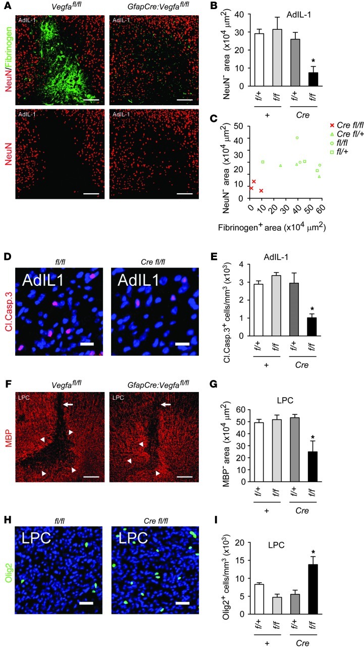
Figure 4. Reduced neuropathology in GfapCre:Vegfafl/fl animals.
(A–C) AdIL-1–injected cortices from 12-week-old GfapCre:Vegfafl/fl mice and littermates (7 dpi, n = 3 per genotype) were (A) stained for NeuN (also shown separately) and fibrinogen, and neuronal loss was quantified and plotted (B) per group and (C) against fibrinogen area (a measure of BBB breakdown). (D and E) Cells from A–C were (D) stained for cleaved caspase-3, and (E) the proportion of cleaved caspase-3–positive cells was determined (a measure of apoptosis). (F–I) GfapCre:Vegfafl/fl mice and littermates (12 weeks old, n = 3 per genotype) received a stereotactic microinjection of lysolecithin into the corpus callosum and sacrificed at 7 dpi. (F and G) Immunostaining and quantification for MBP (a measure of demyelination). Arrowheads, extent of demyelination; arrows, injection tracks. (H and I) Staining and morphometry for Olig2 (a marker of oligodendrocyte loss). Scale bars: 150 μm (A and F); 15 μm (D and H). *P < 0.05, ANOVA plus Bonferroni test. Results are representative of data from 3 independent studies.

