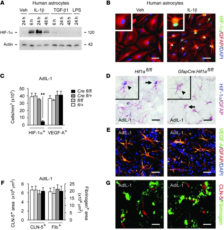
Figure 6. GfapCre:Hif1afl/fl mice show normal VEGF-A expression and BBB opening in inflammatory lesions.
(A) Primary human astrocytes were treated with 10 ng/ml IL-1β, TGF-β1, or LPS for the indicated times, and HIF-1α induction was examined by immunoblot. (B) Human astrocytes were treated with 10 ng/ml IL-1β or vehicle for 24 hours. In IL-1β–treated cultures, HIF-1α localized to astrocytic nuclei (inset; enlarged 2-fold). Results in A and B are typical of data from 3 separate cultures of human astrocytes from different brains. (C–G) AdIL-1 was microinjected into cortical gray matter of 12-week-old GfapCre:Hif1afl/fl mice and littermate controls (n = 4 per genotype), and animals were sacrificed at 7 dpi. Immunostaining and morphometry were performed for (C and D) HIF-1α, (C and E) VEGF-A, and (F and G) fibrinogen (a measure of BBB breakdown) and CLN-5. In D, representative cells (arrows) are shown enlarged 2-fold in the insets (arrowheads). Results in C–G are representative of 3 independent experiments. Scale bars: 30 μm (B, D, and E); 40 μm (G). **P < 0.01, ANOVA plus Bonferroni test.

