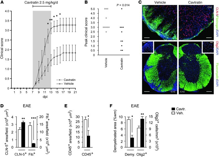
Figure 8. Reduced clinical severity and neuropathology of EAE in cavtratin-treated mice.
Mice (8-week-old male C57BL/6, 10 per group) were sensitized with MOG35–55/CFA, then from onset of weight loss (7 dpi) treated for 7 days with 2.5 mg/kg/d i.p. cavtratin or vehicle. (A and B) Disease was scored using a 5-point paradigm (35) and plotted (A) as a function of time and (B) by peak score. (C) Lumbar spinal cord sections were subjected to immunostaining at 21 dpi to assess BBB breakdown (top) as well as demyelination and oligodendrocyte loss (bottom). (D–F) Morphometry of (D) fibrinogen (a measure of BBB breakdown) and CLN-5, (E) CD45 (a measure of inflammatory cell infiltration), and (F) Olig2 and demyelination (assessed by fluoromyelin). These findings resembled the phenotype of EAE in GfapCre:Vegfafl/fl mice (see Figure 5). Scale bars: 50 μm (C, top); 150 μm (C, bottom); 40 μm (C, insets). *P < 0.05, **P < 0.01, ***P < 0.001, ANOVA plus Bonferroni test (A) or Student’s t test (B and D–F). Data are representative of 3 independent experiments.

