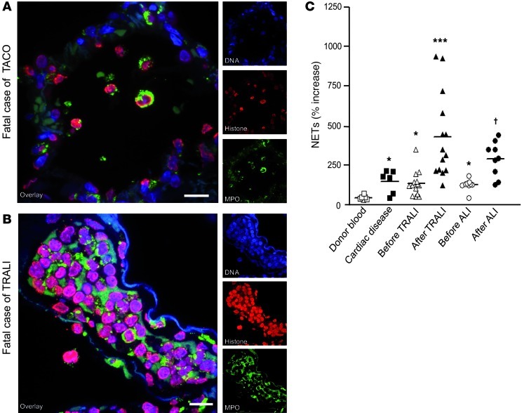
Figure 6. NETs are present in human TRALI lungs and plasma.
(A and B) Human lung paraffin sections were stained for histone (red), MPO (green), and DNA (blue) and analyzed by confocal microscopy. (B) In the TRALI fatality case, we observed clumps of NET-forming neutrophils in the intravascular compartment. (A) In the TACO fatality case, neutrophils were found in mainly intra-alveolar locations, but no NET formation was detected. Scale bar: 10 μm. (C) MPO-DNA ELISA was used to quantify NET components in the plasma of patients, and the mean optical density of plasma obtained from normal, human blood donors (n = 6) was used as the control. Plasma from individuals with cardiac disease (n = 6), individuals before TRALI and after TRALI (paired samples, n = 14), and individuals before ALI and after ALI (paired samples, n = 9) are compared. Horizontal bars represent the mean; symbols represent individual samples. *P < 0.05 versus donor blood group; ***P < 0.001 versus donor blood, cardiac disease, and before TRALI groups; †P < 0.05 versus donor blood, cardiac disease, and before ALI groups.

