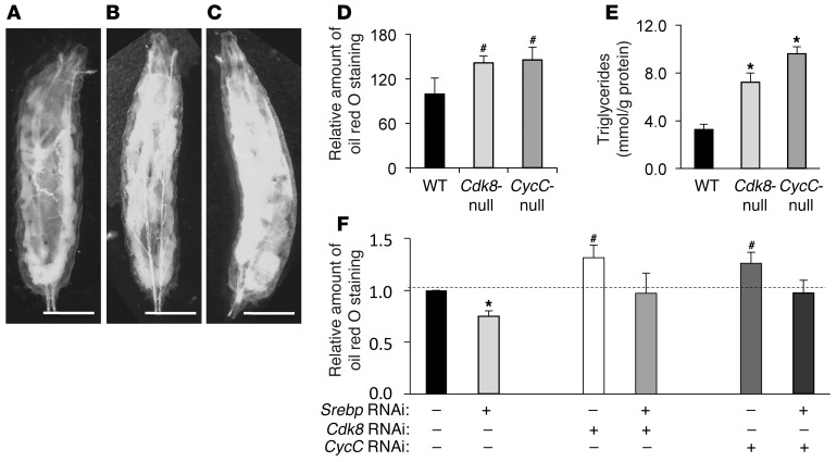
Figure 1. Role of CDK8 and CycC in lipid accumulation in fat bodies of Drosophila larvae.
Representative light micrographic images for larvae (scale bars: 1.0 mm) of (A) wild-type (w1118), (B) Cdk8-null (k185), and (C) CycC-null (y5) Drosophila. (D) Quantitative measurement of oil red O staining in Drosophila larvae of the indicated genotypes. (E) Triglyceride levels in Drosophila larvae of the indicated genotypes. (F) Quantitative measurement of oil red O staining in Drosophila larvae with fat body–specific expression of the indicated RNAi; the background control was FB-Gal4. Data represent mean ± SD of 15 larvae per genotypes for oil red O staining or 3 random pools of the same genotype for triglyceride measurement. *P < 0.01, #P < 0.001 versus control. Data are representative of independent experiments repeated at least 3 times.

