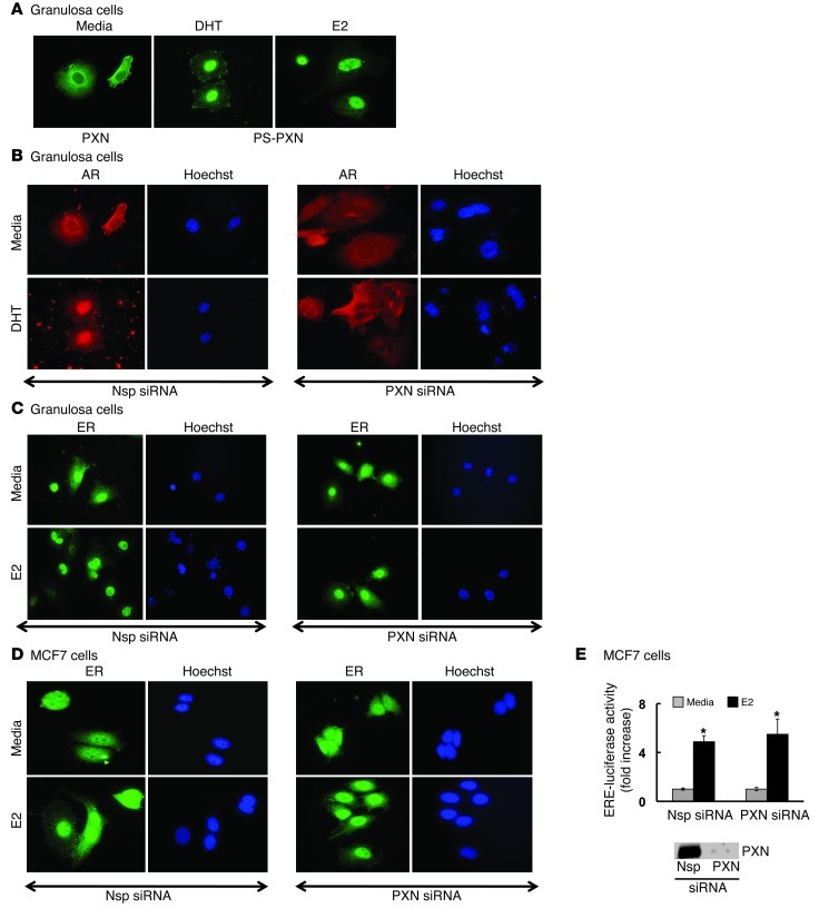
Figure 4. PXN specifically regulates AR nuclear localization in primary GCs.
Primary GCs from C57BL/6J mouse ovaries were treated with DHT or estradiol (25 nM) for 30 minutes. (A) Immunofluorescence studies (n = 3 experiments with identical results) showed that, under basal conditions (medium), PXN is predominantly cytoplasmic. Under DHT or estradiol (E2) stimulation, PS-PXN is primarily nuclear. Adjacent Hoechst staining represents the nucleus. (B and C) Immunofluorescence studies (n = 3 experiments with identical results) of primary GCs treated with Nsp or PXN-specific siRNA. PXN ablation prevents DHT-induced nuclear translocation of AR (B), but has no effect on nuclear localization of ERα in medium or estradiol-treated cells (C). (D) Immunofluorescence studies (n = 3 experiments with identical results) in MCF7 breast cancer cells showing siRNA-mediated knockdown of PXN has no effect on ERα nuclear localization in medium or estradiol-treated cells. Original magnification, ×40. (E) PXN is not required for ERE-mediated transcription. Nsp or PXN-specific siRNA treated MCF7 cells were transiently transfected with ERE reporter luciferase construct plus cytomegalovirus–β-gal plasmid. ERE-luciferase activity was normalized to β-gal expression and data represented as fold increase with respect to medium treatment (mean ± SEM, n = 3). *P ≤ 0.001 relative to medium. The blot on the right shows PXN knockdown.

