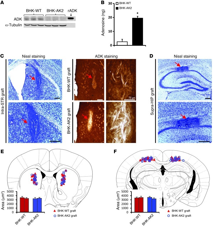
Figure 4. BHK cell–based adenosine augmentation approach.
(A) Representative Western blot from cell extracts of BHK-AK2 (completely lacking ADK expression) and BHK-WT cells (with normal ADK expression, as control) that were used for cell transplantations. Anti-tubulin immunoreactivity was used to normalize for equal loading, and recombinant ADK (rADK) was loaded for comparison. (B) Demonstration of adenosine release from BHK-AK2 cells, which released about 20 ng adenosine per 105 cells during the first hour of incubation. (C and D) Nissl staining of coronal brain sections from graft recipients 3 weeks after grafting into Adk-tg recipients validated the location of the graft (red arrows) in either (C, left) the striatum or (D) above the CA1 pyramidal cell layer of the hippocampal formation. ADK immunohistochemistry (C, right) demonstrated ADK expression in BHK-WT grafts (C, top right) but lack of ADK immunohistochemistry in BHK-AK2 grafts (C, bottom right). The locations of BHK-WT and BHK-AK2 cell grafts of each animal tested are shown superimposed on a standard mouse brain atlas image of the (E) striatum (AP, 1.00 mm from bregma) and (F) hippocampus (AP, –2.10 mm). The graphs by the atlas images indicate the average area of the corresponding cell grafts. Data are mean ± SEM. *P < 0.01, versus BHK-WT. Scale bar: 300 μm.

