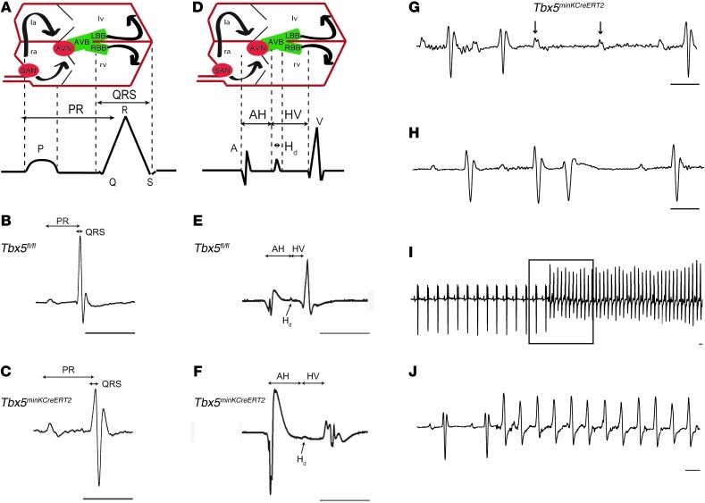
Figure 2. Conduction slowing and arrhythmias after removal of Tbx5 from the ventricular CCS.
(A–J) Conduction system function in Tbx5fl/fl (B and E) and Tbx5minKCreERT2 (C and F–J) mice was evaluated by ambulatory telemetry (B, C, and G–J) and invasive EP studies (E and F). Electroanatomical correlates of ECG and EP recordings are shown in A and D, respectively. la and ra, left and right atria; AVB, AV bundle; AVN and SAN, AV and SA nodes; LBB and RBB, left and right bundle branches. PR and QRS intervals were prolonged during ambulatory telemetry analysis (representative recordings in B and C), and intracardiac recordings (representative recordings in E and F) demonstrated prolongation of AH interval, Hd, and HV interval. Mobitz type II second-degree AV block (G) occurred exclusively in Tbx5minKCreERT2 mice. PVCs (H) were more common in Tbx5minKCreERT2 mice, and ventricular tachycardia (I and J) was observed exclusively in Tbx5minKCreERT2 mice. Boxed area in I is shown at slower scale in J. Scale bars: 50 ms. Arrows in G represent nonconducted p waves. See Table 1 for quantification of ECG and EP intervals.

