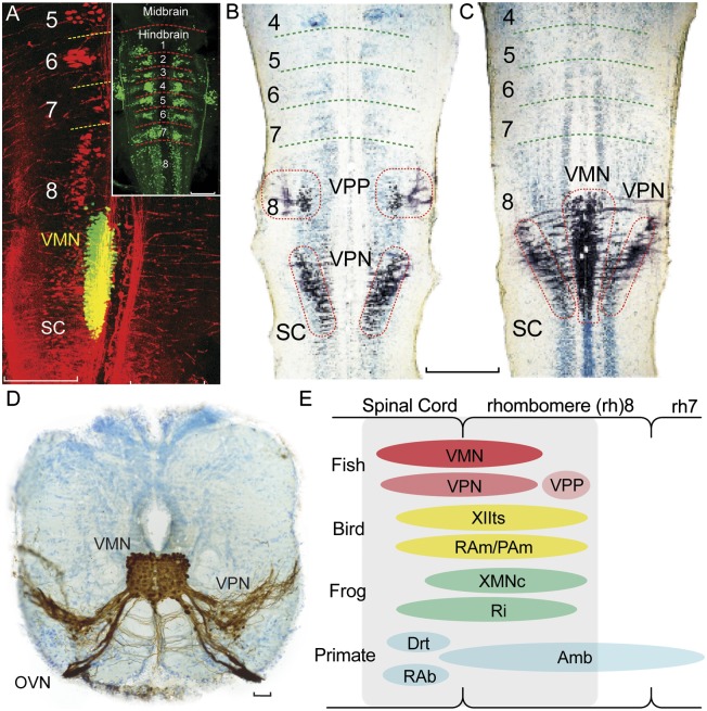
Fig. 3.
Map of developing vocal pattern generator in rh8-spinal compartment. (A) Fluorescently labeled neurons in plainfin midshipman fish larvae visualized with laser scanning confocal microscopy (horizontal plane). Simultaneous visualization of reticulospinal neurons labeled via retrograde transport from the spinal cord (Alexa 546 dextran-amine, red) and VMN (Alexa 488 dextran-amine, green) labeled via the developing vocal muscle. Yellow is composite overlap and does not indicate double labeling. Inset: Clusters of reticulospinal neurons (Alexa biocytin 488, green) in each rh, from 1 to 8. (Scale bars: 0.2 mm.) (B and C) Mapping in horizontal plane of VPP, VPN, and VMN neurons (black) in Gulf toadfish larvae; labeling via transneuronal transport of neurobiotin from developing vocal muscle. Cresyl violet counterstain reveals segmental, reticulospinal clusters. (Scale bar: 0.2 mm.) (D) Transverse section in caudal hindbrain of toadfish showing transneuronal neurobiotin labeling (brown) of paired midline VMN and adjacent VPNs; VMNs and VPNs have extensive dendritic and axonal branching. VMN axons exit via occipital vocal nerve root (OVN; cresyl violet counterstain). (Scale bar: 100 μm.) (E) Sagittal view summarizing relative positions of hindbrain vocal premotor-motor networks in rh8-spinal compartment of fish, birds, frogs, and mammals including primates, based on early-stage and adult phenotypes (see ref. 11 for details). Most laryngeal motor neurons that shape the temporal envelope of mammalian calls originate from caudal nucleus ambiguus (Amb). Drt, dorsal reticular nucleus; PAm, nucleus parambigualis; RAb, nucleus retroambiguus; RAm, nucleus retroambigualis; Ri, inferior reticular formation; XIIts, tracheosyringeal division of hypoglossal motor nucleus; XMNc, caudal XMN. (Adapted from ref. 11.)

