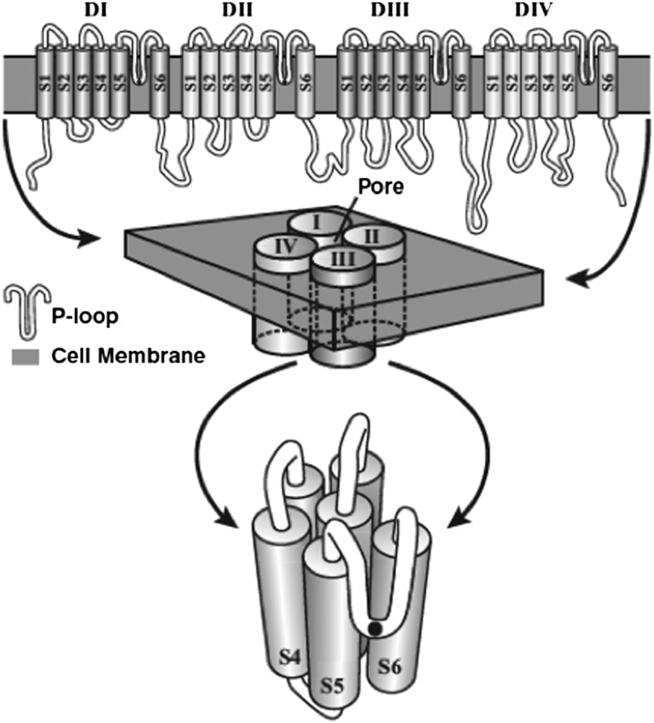
Fig. 2.
Hypothetical secondary structure of a Nav channel. Top: The Nav channel is composed of four repeating domains (I–IV), each of which has six membrane-spanning segments (S1–S6), and their connecting loops (in white). Middle: The four domains cluster around a pore. Bottom: The four P loops dip down into the membrane and line the outer mouth of the channel that is evident in an en face view of a single domain. The black dot represents the single amino acid at the deepest position of each of the four P loops that determines Na+ ion selectivity. From Liebeskind et al. (16).

