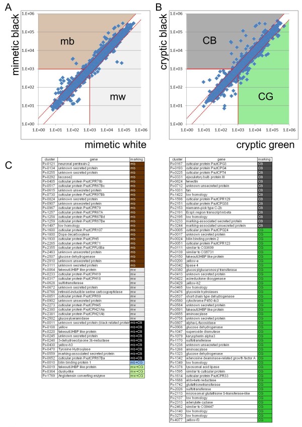
Figure 3.
Screening for marking-specific genes. (A) Scatter plots of normalized signal intensity of mimetic white (x axis, average of six stages) versus mimetic black (y axis, average of six stages). Genes with average intensity more than two-fold (red dashed lines) between mimetic black and mimetic white, and also with an average signal intensity higher than 1,000 (red solid lines) were categorized as marking-specific genes. (B) Scatter plots of normalized signal intensity of cryptic green (x axis, average of five stages) versus cryptic black (y axis, average of five stages). For cryptic markings, T and A were regarded as cryptic green, and E and V were regarded as cryptic black (see Figure 2). (C) List of marking-specific genes. CB: cryptic black-enriched; CG: cryptic green-enriched; mb: mimetic black-enriched; mw: mimetic white-enriched.

