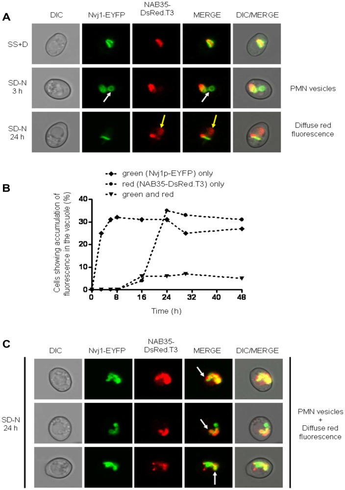
Figure 3. Wild type cells co-expressing Nvj1-EYFP and NAB35-DsRed.T3 nuclear reporters show temporal separation of nucleophagic events.
(A) Wild type (BY4741) cells co-expressing both nuclear reporters were imaged under growing (SS+D) and nitrogen starvation (SD-N) conditions (3 and 24 hours after commencement of nitrogen starvation), respectively. The appearance of Nvj1p-EYFP-derived vesicles (PMN vesicles) in the vacuole is highlighted by white arrows, whereas accumulation of NAB35-DsRed.T3-derived fluorescence (diffuse red fluorescence) is indicated by yellow arrows. (B) Percentage of cells showing accumulation of fluorescence in the vacuole over time: ♦ green (Nvj1p-EYFP) only; • red (NAB35-DsRed.T3) only; ▾ green and red. (C) Accumulation of both Nvj1p-EYFP-derived vesicles (PMN vesicles) and accumulation of NAB35-DsRed.T3-derived diffuse red fluorescence in the same cells 24 hours after commencement of nitrogen starvation. The appearance of vacuolar vesicles containing both nuclear reporters is indicated by white arrows.

