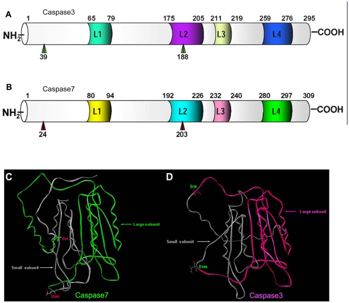
Figure 6. Predicted protein structures of Caspase3 and Caspase7 in Cynops orientalis.
(A)(B) Schematic representation of the predicted subdomain structure of Caspase3 (A) and Caspase7 (B) in Cynops orientalis. The subdomains are shown by the horizontal bar, with the numbers corresponding to the amino acid residues. The cleavage active Aspadine are marked with green or red arrow head, which separate the Caspase3 or Caspase7 into large subunits and small subunits. The four surface loops (L1-L4) that shape the catalytic groove are also labeled with different colors. (C)(D) Cartoon representation of the 3-D structures of Caspase3 and Caspase7. The red color and green stands for the large subunit of Caspase3 and Caspase7, and the gray color indicates the small subunits of Caspase7 and Caspase3. Two cleavage active Asp residues are shown with the form of stick and ball in the cartoon. The 3-D structure prediction was performed by I-TASSER server.

