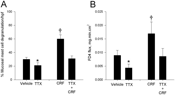
Figure 8. The Role of the enteric nervous system in CRF-mediated mast cell activation and FD4 flux.
Toluidine blue staining of histological sections (100 X magnification) (A) and quantitation of the % of degranulated tissue mast cells (B) in porcine ileum revealed increased degranulation of mast cells in CRF–treated tissues. Treatment of ileal tissues with tetrodotoxin (TTX) inhibited mast cell degranulation and FD4 flux (C) under basal and CRF-stimulated conditions and inhibited. For histological analysis, values represent means ± SE; n = 6 animals and are presented as the percentage of total cell count per treatment at 20× magnification. FD4 flux values represent means ± SE; n = 6−8 animals Symbols (*,†) differs significantly by p<0.05 from vehicle control.

