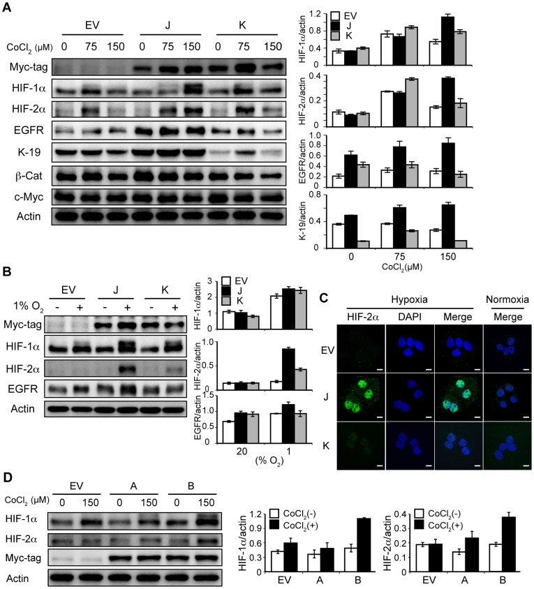
Figure 4. Loss of the SxxSS motif in TCF-4 isoforms increases expression of HIF-αs under hypoxia.
(A and B) Immunoblot analysis of empty vector-transfected (EV), TCF-4J-overexpressing (J), and TCF-4K-overexpressing cells (K). The cells were treated with 0 and 150 µM CoCl2 (A) or cultured under 1% oxygen tension for 48 hr (B). Cell lysates were subjected to detect HIF-αs and Myc-tag TCF-4 isoform protein expression. Expression levels were plotted as a ratio to actin (right panel). (C) Confocal microscopy for nuclear localization of HIF-2α protein. Cells were treated with 0 µM (Normoxia) or 150 µM (Hypoxia) CoCl2 and stained with an anti-HIF-2α antibody (green). DAPI was employed for nuclear staining. (D) Immunoblot analysis of total cell lysates from cells treated with 0 or 150 µM CoCl2 for 48 hr. A and B represent HAK-1A cells overexpressing TCF-4A (“short form” of TCF-4K) and TCF-4B (“short form” of TCF-4J), respectively (see Figure 2A). Note the robust increase in expressions of HIF-1α and HIF-2α in B cells.

