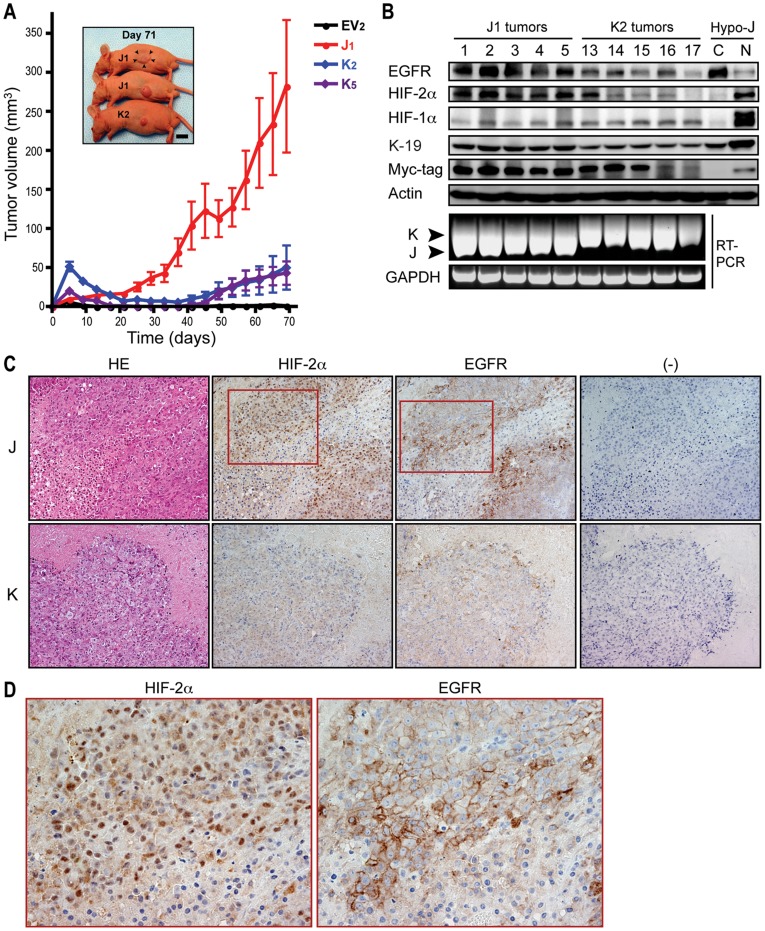
Figure 6. Loss of the SxxSS motif in TCF-4 isoforms promotes tumorigenesis.
(A) Representative experiment demonstrating xenograft tumor development and growth rate. EV2, control; J1, J cell; K2 and K5, K cell clones. (B) Protein expression of the indicated molecules in J1 tumors (1–5) and K2 tumors (13–17) by immunoblot analysis. Nuclear (N) and cytoplasmic (C) proteins expressed in the 150 µM CoCl2-treated J cells (Hypo-J) were used as positive controls. (Bottom panel) TCF-4J and K mRNA expression was verified by RT-PCR in J1 and K2 tumors; GAPDH, glyceraldehyde-3-phosphate dehydrogenase. (C) Immunohistochemistry for HIF-2α and EGFR expression in representative J- and K-cell derived tumors. Original magnification, 200x. HE, hematoxylin and eosin; and (-), negative staining. (D) Magnified view (400x) for the corresponding squared areas in (C) for HIF-2α and EGFR expression.

