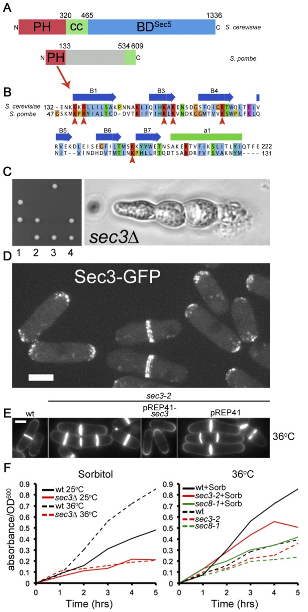
Figure 1. Function and localization of S. pombe Sec3.

A. Scheme of Sec3 in S. cerevisiae and S. pombe. Cryptic PH domains are shown in red, predicted coils in green. The C-terminal Sec5-binding region of S. cerevisiae Sec3 [25] is not conserved in S. pombe. B. Alignment of Sec3 PH domain. Critical residues predicted to contact phospholipids are indicated with red arrowheads and are conserved in S. pombe. C. Tetrad dissection of sec3Δ::kanMX/sec3+ diploids on YE plate and terminal multi-septated phenotype of unviable spore. None of the viable spores grew on G418 plates (not shown). D. Maximum projection of spinning disk confocal sections of Sec3-GFP. Bar is 5 µm. E. Calcofluor-stained wildtype and sec3-2 cells grown at 36°C for 6 h. Note that the multi-septated phenotype of sec3-2 is rescued by plasmid-expression of sec3+ (pREP41-sec3+), but not empty vector (pREP41). Bar is 5 µm. F. Secretion of acid phosphatase in wildtype and sec3 mutants. Left: wildtype and sec3Δ mutants were pre-grown in EMM 1 M sorbitol at 25°C and kept at 25°C or shifted to 36°C at t = 0. Right: wildtype, sec3-2 and sec8-1 mutants were pre-grown at 25°C in EMM 1 M sorbitol and shifted to 36°C ± sorbitol at t = 0.
