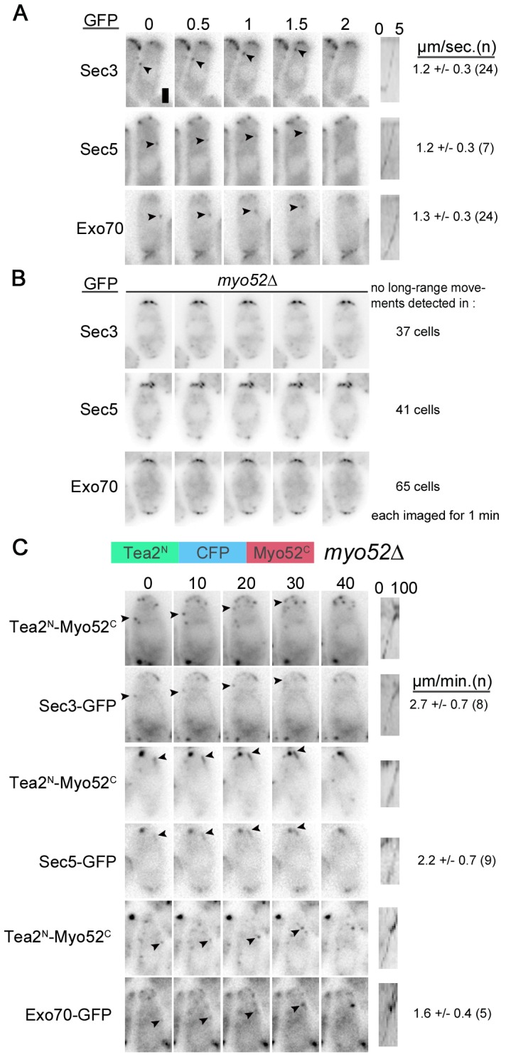
Figure 5. Sec3, Exo70 and Sec5 are transported towards cell poles by myosin V Myo52.

A-B. Timelapse images of Sec3-, Sec5- and Exo70-GFP in wildtype (A) and myo52Δ cells (B). Arrowheads point to moving dots. A kymograph along the path of the indicated dot is shown on the right. Average rate of movement is shown on the right. Time is indicated on top in seconds. C. Timelapse images of Sec3-, Sec5- and Exo70-GFP in myo52Δ cells expressing a Tea2N-CFP-Myo52C chimera. GFP and CFP signals are shown. Arrowheads point to moving exocyst dots, which colocalize with the motor chimera. Note other non-moving signal also colocalize. A kymograph along the path of the indicated dot is shown on the right. Average rate of movement is shown on the right. Time is indicated on top in seconds. All images are single widefield images. Bar is 2 µm.
