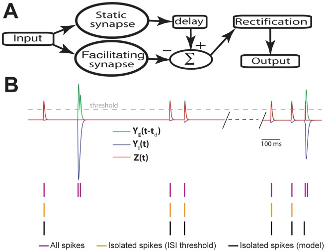
Figure 10. A biophysically plausible neural circuit can accurately extract isolated spikes and therefore decode their information about movement direction.
A) Schematic of the decoding model for isolated spikes. It consists of parallel processing by two synapses with one displaying facilitation and the other displaying no plasticity (i.e. “static”). The output from the static synapse YE(t) is delayed and the output from the facilitating synapse YI(t) is then subtracted from it. This signal is then half-wave rectified to give the output Z(t). Finally, Z(t) is thresholded to obtain the output spikes. B) Performance of the decoding model compared with the performance of an ISI threshold criterion at detecting isolated spikes. Shown are the delayed output of the static excitatory synapse YE(t−td) (green trace), facilitating inhibitory synapse YI(t) (blue trace), and the output of the model Z(t) (red trace) with the threshold used to detect output spikes (dashed gray trace), the original spike train (purple ticks), the isolated spikes according to the ISI threshold (orange ticks), and the isolated spikes according to the decoding model (black ticks). Parameter values used were τF = 200 msec, τD = 500 msec, τE = 5 msec, τI = 8 msec, GI = 5, I0 = 3.41 msec, td = 4 msec.

