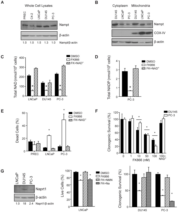
Figure 1. Nampt regulates NAD+ levels and survival in PCa cells.
(A) Nampt protein levels were determined by western blotting in PREC and PCa cells. (B) Nampt localization was also determined in the cytoplasmic and mitochondrial fractions of PCa cells by western blot, with COX-IV as a mitochondrial marker (C) Total NAD levels were measured in PCa cells after 48 hour treatment with vehicle (DMSO 0.1%), or FK866 (100 nM) in the absence or presence of NAD+ (100 µM) (*p<0.01). (D) Total NADP levels were measured in PC-3 cells after 48 hour treatment with vehicle (DMSO 0.1%), or FK866 (100 nM) in the absence or presence of NAD+ (100 µM) (*p<0.01). (E) The effect of FK866 on survival was assessed by trypan blue exclusion in PREC, LNCaP, and PC-3 cells after 48 hours of treatment. (F) Clonogenic survival of PC-3 and DU145 cells was measured following a dose-response of FK866, or 100 nM FK866 plus NAD+ (100 µM) for 24 hours (*p<0.01). (G) Expression of Naprt1 and β-actin were determined by western blot. The ability of NMN and Na to protect cells from FK866 was determined by trypan blue exclusion in LNCaP cells (48 hour treatment) and clonogenic survival in PC-3 and DU145 cells (24 hour treatment).

